Over 40 Years of Dedicated Service
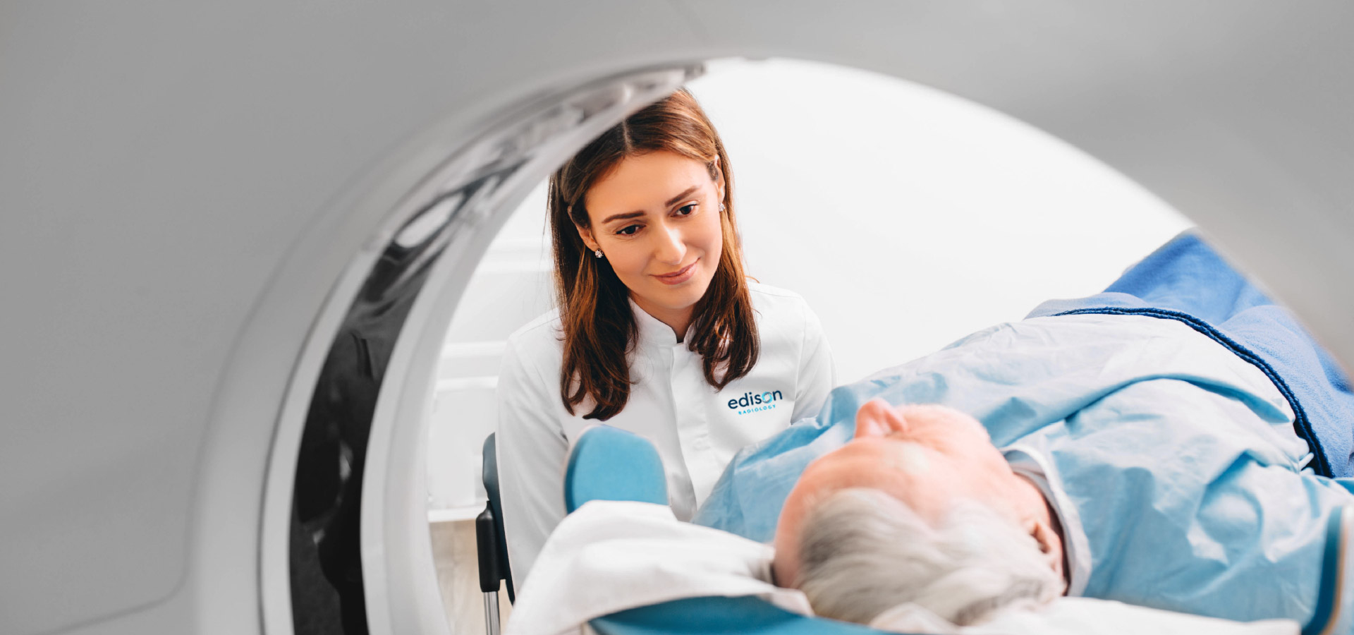
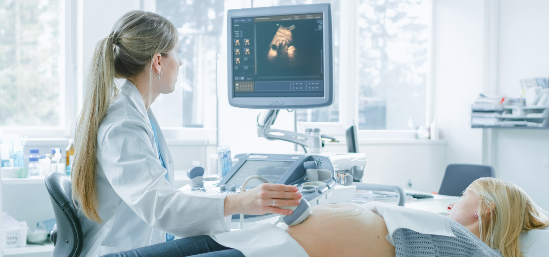
Edison Radiology Group is a leading provider of diagnostic and interventional radiology services in Central New Jersey. We utilize the most advanced imaging services and equipment, providing academic level quality with compassionate care. We provide continuous diagnostic and interventional radiology services to JFK Medical Center and Muhlenberg Medical Center, part of Hackensack Meridian Health and operate three outpatient multi-modality imaging centers in Edison, Old Bridge and Toms River.
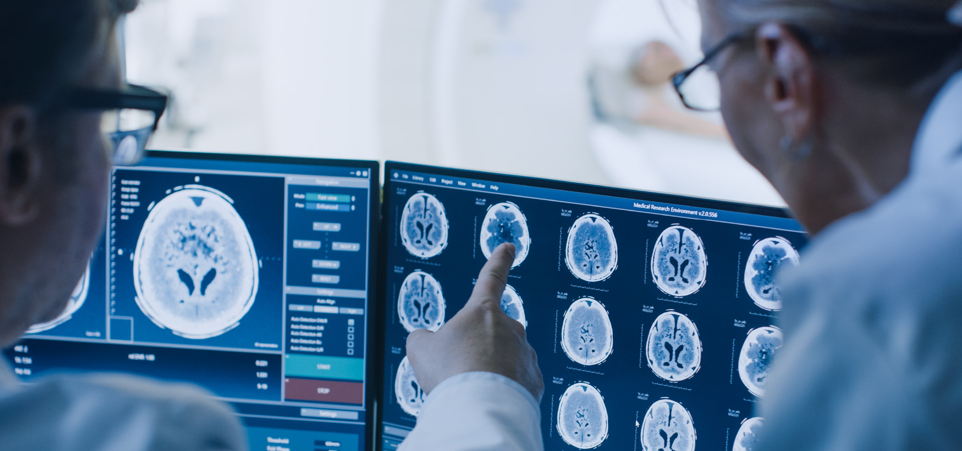
All of our radiologists are board certified by the American Board of Radiology and most have sub-specialty fellowship training in their areas of expertise. They are supported by our highly talented and certified technologists who also have specialized training in their specific fields.
Our referring physicians trust us for our expertise and our patients appreciate us for our compassionate care. We are an academic practice in your backyard!
From chest radiographs to complex cancer screening, from MRs to interventional radiology surgeries, we are your go to radiology group in central New Jersey.